


Поделиться случаем QuikVue Vet - разрешение абсцесса роговицы
We are glad to share a vet case study captured by QuikVue eye imaging adaptor from Dr. Allison Fuchs.
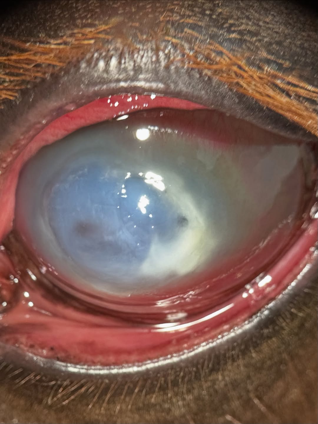
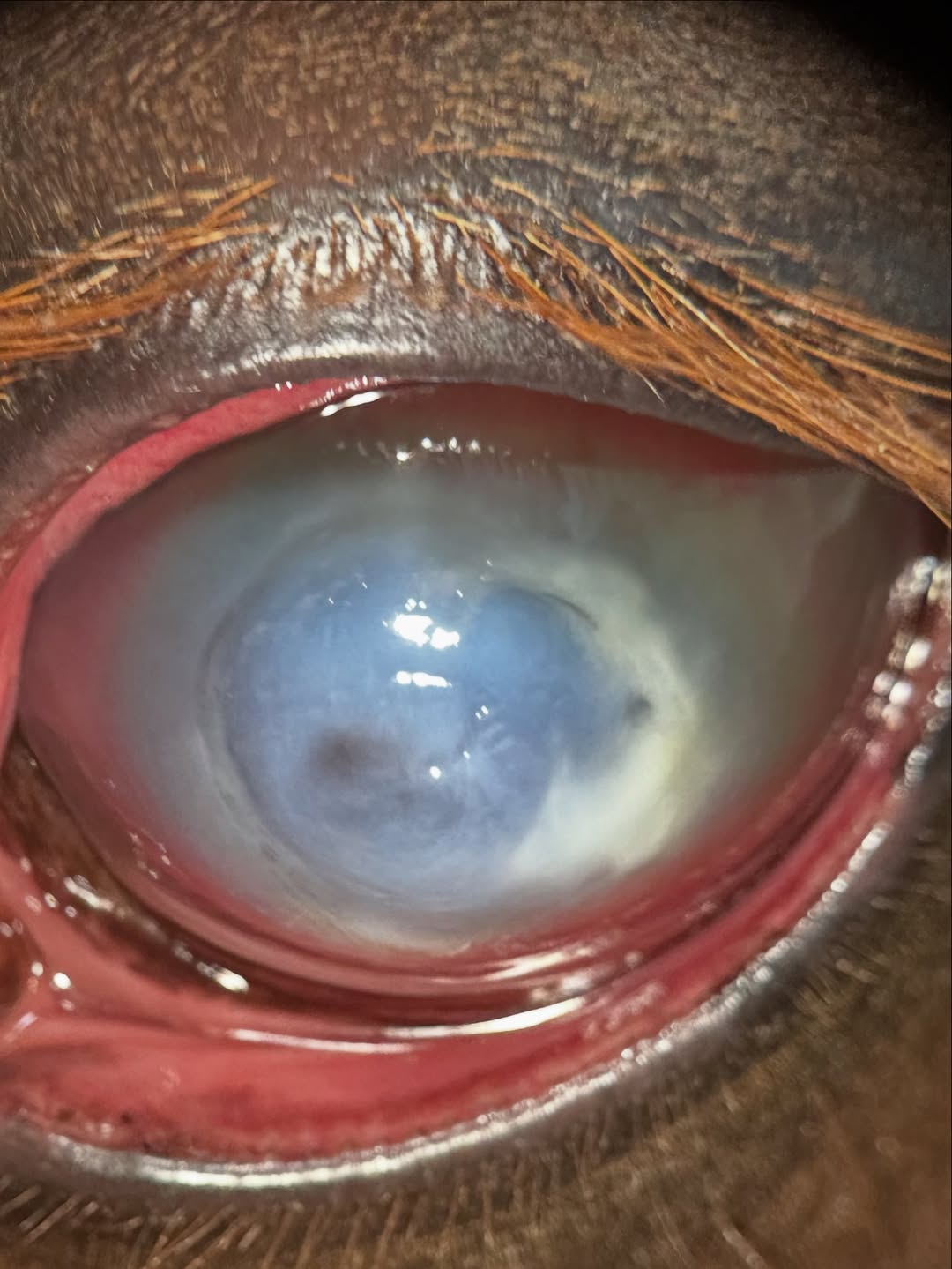
This horse was initially seen about a month ago and diagnosed with a corneal abscess, as seen in the first two photos. Basically a nasty corneal ulcer that became infected, and sealed over with bacteria and/or fungus trapped in the deeper layers of the cornea. This is unfortunately common in horses and can be really difficult to treat.
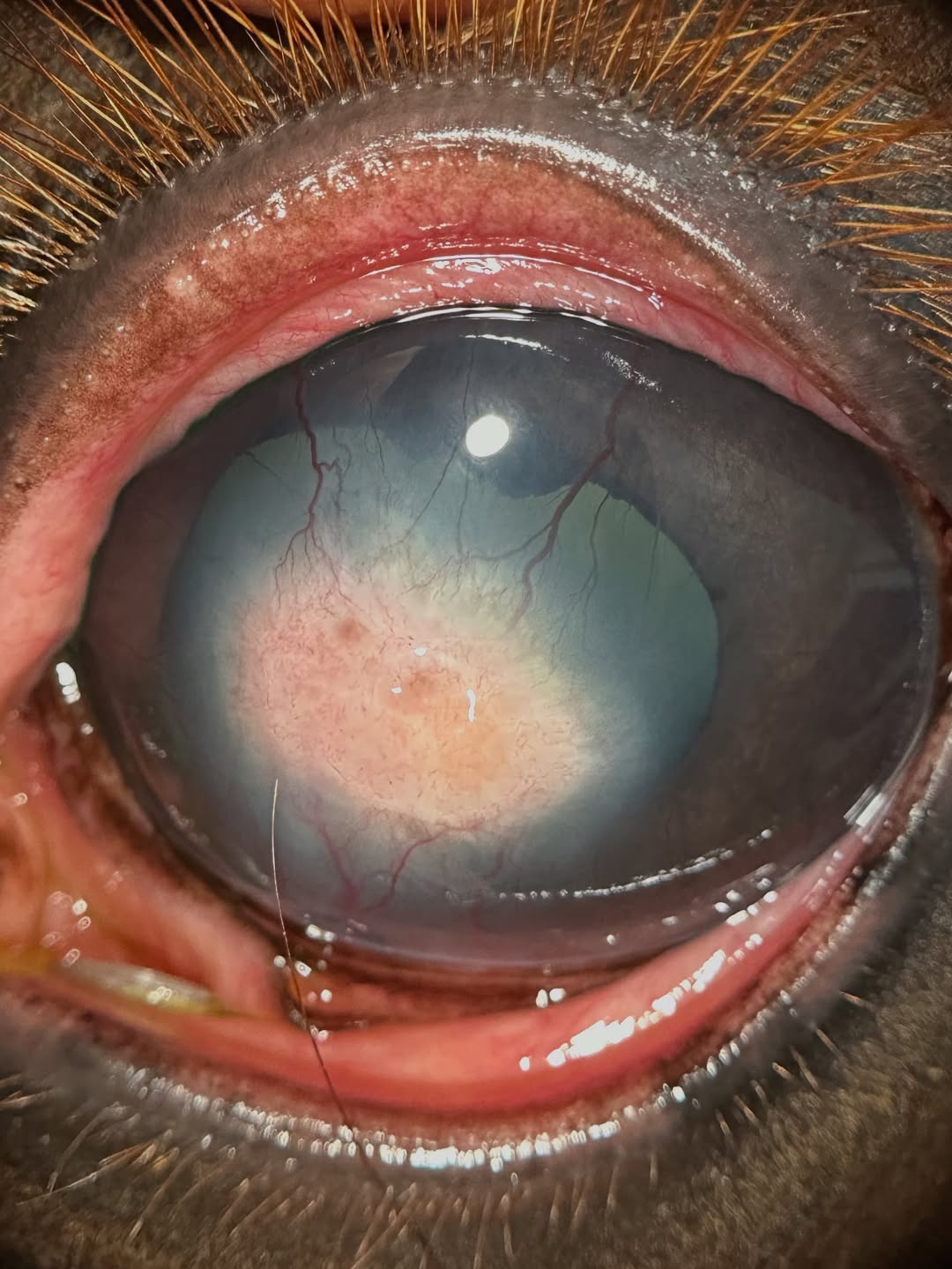
Fortunately we were able to treat this young mare aggressively, and in the third photo she is completely healed. There is an area of scarring and granulation tissue in the middle of the cornea, which is the pinkish spot, but that scar will fade over the next few months. She can see around it, and should end up with pretty normal function long term!
 |  |  |
We are glad to share a vet case study captured by QuikVue eye imaging adaptor from Dr. Allison Fuchs.
This horse was initially seen about a month ago and diagnosed with a corneal abscess, as seen in the first two photos. Basically a nasty corneal ulcer that became infected, and sealed over with bacteria and/or fungus trapped in the deeper layers of the cornea. This is unfortunately common in horses and can be really difficult to treat.
Fortunately we were able to treat this young mare aggressively, and in the third photo she is completely healed. There is an area of scarring and granulation tissue in the middle of the cornea, which is the pinkish spot, but that scar will fade over the next few months. She can see around it, and should end up with pretty normal function long term!
 |  |  |


